Biology
- Human Body Systems
Human Body Systems!Integumentary System: Your body is very prone to damage, and that's why we have this system! This is an organ system that helps protect the body. It has many functions such as to cushion, excrete wastes, regulate temperatures...
- Colon
Term: colon Origin: An Greek ?????/colon(=large intestine) > the word ????? is the pariciple of the verb ??????/kolazo (=punish, excaudate, cut) Definition: The last part of the digestive system that moves waste material from...
- Pancreas
Term: pancreas Origin: Anc Greek ???/pan(=all) + ?????/creas(=flesh, meat) presumably because of its fleshy consistency as this organ contains neither cartilage nor bone. Coined : The pancreas was first identified for western civilization by Herophilus...
- Organs Derived From Germ Layers Of Embryo
Ectoderm Mesoderm Endoderm Skin (epidermis) and their pigment cells, hairs, nails. Connective tissues, superficial and deep fascia, ligaments, tendons, dermis of skin (from dermatotome) Epithelial part of mouth, some part of palate, tongue,...
- Respiratory System.
Respiratory System.The cells of our body consume oxygen in order to obtain energy. In this process, called oxidation, the cells burn glucose using oxygen, releasing carbon dioxide, and obtaining the necessary energy to carry out several metabolic processes....
Biology
Digestive System
Digestive System.
 |
| Kinder beim Mittagessen, M.L.Benjamin Vautier |
The digestive system is the main one responsible for processing the food we consume, so that it can be used as an energetic support or as a font of source materials.
The digestive system fragments large molecules of the food transforming them into smaller molecules that can be absorbed. In this unit we will analyse the anatomy, physiology and reproduction of eukaryotic animal cells.
The whole digestive process can be divided into five consecutive actions.
- Ingestion: the food passes from the exterior to the interior of the body via the mouth.
- Movement of the food: the food moves across the digestive tube.
- Digestion: fragmentation of the food by mechanical and chemical processes.
- Absorption: the nutrients obtained when the food is digested are transported from the gastrointestinal duct to the blood or lymphatic system.
- Defecation: release of non-digested substances.
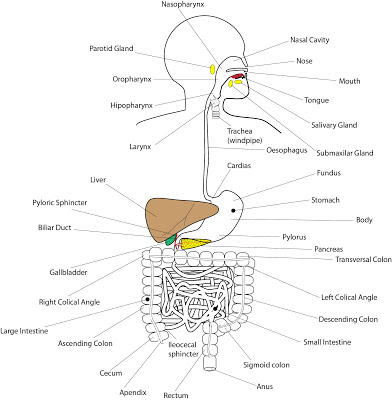
Anatomy of the Digestive System.
Introduction.
The digestive system can be divided into two parts:
- Gastrointestinal duct: it is a nine meter long tube that starts in the mouth, crosses the thorax and abdomen and ends at the anus.
- Accessory structures: there are several different structures. The most frequent ones are glands, although there are other structures such as teeth or the tongue.
In this unit we will study the digestive duct, from the mouth to the anus, analysing the most relevant accessory structures in each part.
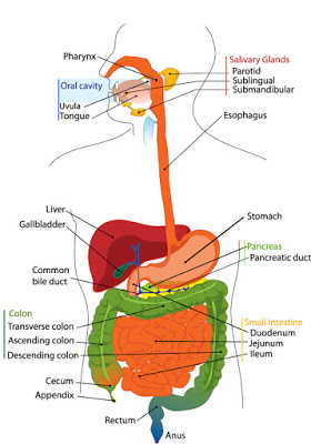
Mouth.
Also called oral cavity, this is the gate where the ingestion of food takes place. It is bound laterally by the cheeks, at the right and left sides, the hard and soft palates at top and the tongue at floor. It is separated from the exterior by two fleshy folders covered by thin skin called lips.
The inner part, between the cheeks is called vestibule. Behind the vestibule, the duct that binds the mouth posteriorly is called isthmus of the fauces. This duct separates the mouth from the pharynx. Just in the boundary of the isthmus there is a little hanging piece of soft tissue called uvula.
The roof of the mouth is called the palate and has two parts. The anterior part is made of bone and it is called hard palate. The bones that form the hard palate are two small palatine bones and a part of the maxillary bone. The posterior part is made of muscle and it is called soft palate.
There are also several accessory structures in the mouth:
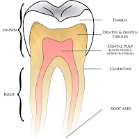 |
| Tooth Anatomy |
- Tongue: this is a skeletal muscle that forms the dynamic structure of the mouth floor. It is essential to break the food down using the teeth and then to swallow the bolus. The taste sensor is also located on the surface.
- Salivary Glands: they constantly secrete saliva to the mouth. So that they wet the oral cavity and the pharynx. When the food enters into the mouth the secretion rises. Saliva lubricates the food and dissolves part of its components, starting the chemical digestion of several components. There are three pairs of salivary glands. One pair are called Parotids and they are located beneath the ears. Another pair of glands are called Submandibular and they are located in the posterior part of the mouth floor, just beneath the tongue base. The last pair of glands are called Sublingual and they are located in the anterior part of the mouth floor, under the central part of the mouth.
- Teeth: accessory structures located in the alveolar apophysis of the maxillary bones and mandibular bone, which are covered by the gums. They are very hard and resistant. When the food is chewed, the teeth cut and crush the food. Adult human beings have 32 teeth. There are four types of teeth and adult human beings have 8 incisors, 4 canines, 8 premolars and 12 molars.
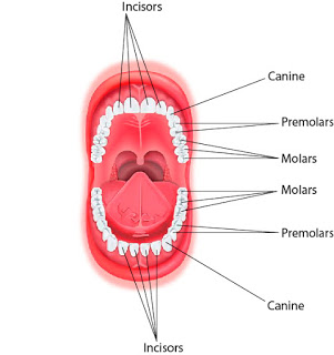 |
| Mouth: Teeth. |
Pharynx.
This duct is a part of the digestive and respiratory systems at the same time. The contraction of the muscles that surrounds the tube moves the food from the mouth to the oesophagus. It is in contact with upper part of the larynx, called epiglottis, that prevents the food from passing into the respiratory system.
Oesophagus.
This is a 20 to 30 centimetre long tube, located beneath the windpipe (trachea). It starts at the lower portion of the pharynx, crosses the mediastinum trespassing the diaphragm through the oesophagic hiatus and ends up at the upper portion of the stomach.
The peristaltic movements of the wall transport the bolus from the pharynx to the stomach. The tube terminates at the cardias, also called superior oesophageal sphincter, that controls the entrance to the stomach.
Stomach.
Dilated portion of the gastrointestinal duct, located under the diaphragm. Its shape and size changes according to the movements of the diaphragm, the amount of food and the digestion phase. It has four parts. The upper part, called cardia, has the opening to the oesophagus. Just under the cardias, at the left side, there is a large concave part called fundus. The central part of the stomach is called body. And the lower part, connected to the intestine by a sphincter called pylori, is called pyloric antrum.
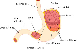 |
| Stomach Antomy. |
Cardia and pylori are two sphincters stat enclose the food in the stomach during the digestive process of the stomach, so that the food being digested and the digestive acids can not flow back to the oesophagus or pass into the intestine until the process has finished.
Two important digestive processes take place in the stomach: mechanical digestion and chemical digestion. The mechanical digestion is carried out due to the peristaltic movements of the stomach wall. These movements also mix the food with the gastric juices, released by the stomach wall. These juices are responsible for the chemical digestion of the food. The main component of the liquid that carries out the chemical degradation of food components is the hydrochloride acid (HCl), although there are other digestive components, such us enzymes responsible for degradation of proteins, fats or polysaccharids. The bolus after being digested in the stomach is called chyme.
Small Intestine.
It is a six meter long and three centimetre thick tube, that stats at the pyloric sphincter, just beneath the stomach and ends up at the ileococcal sphincter, that separates the small intestine from the large intestine. It is intricately tingled in the central and inferior region of the abdominal cavity.
The small intestine has three parts. The first one is called duodenum, it is only 25 centimetre long and it is located just beneath the stomach. It is an important region, because liver and pancreas release their secretion to it. The central part of the small intestine is called jejune and it is 2.5 meter long. The final part is between 3.5 and 4 meter long and it is called ileum. The ileocecal sphincter separates the ileum from the large intestine.
The small intestine wall is not smooth, but it is profusely folded, forming lots of little finger shaped protrusions called intestinal villi. Their function is increasing the intestinal surface, so intestine has an extremely huge area from which to absorb nutrients from the food. The epithelial cells that cover the inner part of the tube, besides, have microvilli in their upper membrane, to increase the absorptive surface even more.
As we have mentioned, the absorption of nutrients takes place in this part of the digestive system. In fact, it is the only place where a massive absorption takes place. And it is also the place where the digestion of food is completed, although it has started in the mouth and above all in the stomach. The mechanical digestion in the small intestine, led from the peristaltic movements of the wall, is not really relevant. The chemical digestion, however, is very important. It transforms the chyme from the stomach into chyle. The cells of the intestinal wall release little amounts of digestive products, although chemical digestion is principally carried out by chemical substances released to the intestine by two accessory glands: pancreas and liver.
- Pancreas: this is a 12.5 centimetre long and 2.5 centimetre thick (although the thicker part is usually slightly thicker) gland. It is located beneath the major curvature of the stomach. It is connected to the small intestine by two ducts. The gland has two parts, the thicker part near duodenum and called the head, and the thinner part, slightly sharp called the tail. The pancreas has two main functions. On the one hand, pancreas produces and releases pancreatic juice, a liquid rich in enzymes responsible for degradation of nutrients: amylases that degrade sugars, proteases that degrade proteins and lipases that degrade lipids. On the other hand, it produces and releases digestive hormones that control the levels of glucose in the blood. This function is carried out by a section of the pancreas called endocrine pancreas. The pancreatic duct that connects the pancreas with the intestine joins with the bile duct from the gallbladder.
- Liver: this is the largest gland of our body. It weighs around 1.5 kilogrames. It is located under the diaphragm, taking up a big space of the right hypocardiac and a part of the abdominal epigastric. It is divided into two big lobules, called left and right lobule, joined together by the falciform ligament. It is irrigated by the hepatic arteries, that provide oxygenated blood, and by the inferior vena cava, that arrives from the small intestine and provides nutrients. The liver produces and releases between 800 and 1000ml of gall per day. This pale green liquid is responsible for increasing the pH of the food that has been digested n the stomach, and for promoting the emulsion of lipids and fats, so that large a cumulus of fats are transformed into little drops of lipids that can be easily absorbed. The gall is also used to release waste products produced by the liver, such as bilirubin, made from destroyed erythrocytes and responsible for the brown colour of the faeces. Gall is produced by the liver, but it is not directly released to the intestine, but is stored in the gallbladder.
- Gallbladder: it is a pear-shaped sac, where gall is stored. It is located just under the lower depression of the liver. The bile duct connects the gallbladder with the pancreatic duct, and both ducts joined connect with the intestine.
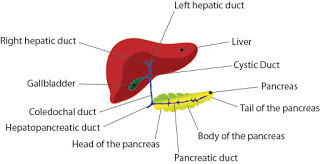 |
| Anatomy of liver, gallbladder and pancreas. |
Large Intestine.
This is a 1.5 meter long and 6 meter thick tube, that starts at the cecum and ends up in the anus. Large and small intestine are separated by the ileocecal sphincter. There is a 6cm long section of tube just below the sphincter, called cecum. At the end of the cecum a little cylindrical structure called vermiform appendix can be found. The appendix and cecum are the right side of the abdomen. The rest of the large intestine is called colon and can be divided into several parts. First, the ascending colon from the ileocecal sphincter to the right-upper part of the abdomen, just beneath the diaphragm. Then the colon turns 90° in the right colical angle. The portion of colon that crosses the abdomen from the right to the left side of the abdomen, parallel to the diaphragm axis, is called transverse colon. It turns 90° again in the left colical angle, and crosses the abdomen from the left-upper to the left-lower part of the abdomen. It is called descendant colon. The descendant colon turns 90° again just when it is parallel to the iliac crest, and then is called sigmoid colon. The tube turns again and this final part is called rectum. In the last 3cm of the rectum there is a large sphincter called anus, that controls the release o faeces to the exterior. The colon is the place where non digested and non absorbed substances arrive. There are many bacteria that transform these into waste product. Some vitamins are produced by these bacteria, and can be absorbed by large intestine. Waste products, however, are not digested or absorbed, only a large amount of water from the faeces is absorbed, to prevent our body from dehydration. So that the faeces produced after a correct digestive process are nearly dry, and made up of substances that our body can not digest or absorb.
 |
| Digestive System. |
Physiology of the digestive system.
The whole digestive process is based on the physical and chemical degradation of the food in order to transform the complex molecules into small and simple ones that could be absorbed and transported from the interior of the small intestine to the blood or the lymph. Non digested or absorbed substances must be dried, condensed and released to the exterior.
This digestive process starts in the mouth. Teeth cut the food in small pieces and some chemical components of saliva carry out the first chemical digestive step. Salivary amylase degrades complex sugars, for instance, transforming them into smaller and simpler sugars. The food, after being chewed and mixed with saliva is called bolus.
After being swallowed the bolus descends along the oesophagus, arriving at the stomach. The bolus is enclosed in the stomach, because two sphincters, cardiac and pylori, are closed. So that the bolus cannot pass to the intestine until it has been digested, but cannot either flow back to the oesophagus.
The stomach wall releases extremely acidic gastric juices, rich in degradative enzymes that destroy large molecules, transforming them into smaller ones, such as the enzyme called Pepsine, that breaks down proteins transforming them into simple amino acids. The stomach wall, besides, carries out abrupt peristaltic movements that provoke the mechanical digestion of food and improve the chemical digestion mixing the food with the gastric juice. The food after being digested by the stomach is called chyme.
The final chemical and physical digestive process of the food takes place in the small intestine. The chemical digestion in the small intestine is carried out by the pancreatic juice. This liquid is rich in degradative enzymes: proteases that destroy proteins transforming them into amino acids, lipases that destroy fats transforming them into simple lipids and amilases that destroy polysaccharides transforming them into simple sugars. Pancreatic juice and gall also neutralise the pH of the chyme, which is very acid due to the hydrochloride acid of the stomach. Emulsification of fats is another important function of gall. This emulsification is essential to improve the absorption of lipids and fats by the small intestine.
The food after being digested in the small intestine is called chyle. And chyle is the final product ready to be absorbed by the small intestine. As we studied before, the inner wall of the intestine is profusely folded to increase the surface in contact with the food, improving the absorption of nutrients. The exterior of the small intestine is highly vascularised, so that the nutrients are transported directly from the inner part of the intestine to the blood vessels and lymphatic system. The transportation of nutrients is a controlled process and only small digested substances, such as simple sugars, amino acids or small fats are able to trespass the barrier. The fats are mainly transported by the lymphatic system, whereas the rest of nutrients are transported by the circulatory system.
Large intestine hardly absorbs any nutrient, only some vitamins released by bacteria and, above all, large amounts of water. The inner surface of the large intestine is smooth, with no folds (merely because they are not necessary). The absorption of water is extremely important, preventing our body from dehydration.
After this process, all the dehydrated remains, made up of non digested and non absorbed substances reach the end of the tube and are released, during the defecation, through the anus.
As we studied, liver is responsible for provision of gall, but it has also other important digestive functions. First, it is responsible for storing some nutrients, such us sugars (forming a macromolecule made of large chains of sugar called glycogen) and fats. Second, liver transforms toxic products, such as cholesterol and bilirubin and releases them into the intestine, so that they are expelled them to the exterior. And third, liver is responsible for many metabolic reactions, such us gluconeogenesis, a group of chemical reactions essential to produce glucose from other simpler metabolic products.
The pancreas is very important to control some digestive processes. It produces two hormones, insulin and glucagon, that control the glucose balance. Insulin is produced when the glucose level rises, promoting actions to reduce it. On the other hand glucagon is produced when the glucose level drops, promoting actions to increase it. This actions are very important, because the glucose level in blood must stay relatively constant.
 |
| Digestive System |
- Human Body Systems
Human Body Systems!Integumentary System: Your body is very prone to damage, and that's why we have this system! This is an organ system that helps protect the body. It has many functions such as to cushion, excrete wastes, regulate temperatures...
- Colon
Term: colon Origin: An Greek ?????/colon(=large intestine) > the word ????? is the pariciple of the verb ??????/kolazo (=punish, excaudate, cut) Definition: The last part of the digestive system that moves waste material from...
- Pancreas
Term: pancreas Origin: Anc Greek ???/pan(=all) + ?????/creas(=flesh, meat) presumably because of its fleshy consistency as this organ contains neither cartilage nor bone. Coined : The pancreas was first identified for western civilization by Herophilus...
- Organs Derived From Germ Layers Of Embryo
Ectoderm Mesoderm Endoderm Skin (epidermis) and their pigment cells, hairs, nails. Connective tissues, superficial and deep fascia, ligaments, tendons, dermis of skin (from dermatotome) Epithelial part of mouth, some part of palate, tongue,...
- Respiratory System.
Respiratory System.The cells of our body consume oxygen in order to obtain energy. In this process, called oxidation, the cells burn glucose using oxygen, releasing carbon dioxide, and obtaining the necessary energy to carry out several metabolic processes....