Biology
- #84 Question 7
Beetroot cells contain a red pigment that cannot normally escape from the cells through the cell surface membrane. A student carried out an investigation into the effect of temperature on the permeability of the cell surface membrane of beetroot cells....
- #82 Question 5
The diagram below shows a small part of a human lung as it appears through a microscope. (a) Name the type of blood vessel in which the red blood cell is present. (1 mark)(b) Describe and explain two ways in which the structure of the alveoli, shown...
- #80 Question 3
(a) The diagrams show a cell in various stages ofthe mitotic cell cycle. Name the stage represented by each diagram, and arrange them in the correct sequence. (b) Describe the role of spindle microtubules in mitosis. (3 marks) (c) The graph below shows...
- #78 Question 1
(a) The diagram shows a small part of a cell, as seen using an electron microscope. (i) Name the parts labelled A to D. (2 marks) (ii) Describe how part B is involved in the formation of extracellular...
- Prokaryote: Cells
In this picture are millions and millions of bacteria. No matter how much I clean the countertops, once an object or hand is placed on it, more bacteria live there. Bacteria is a prokaryote which are cells that do not have a nucleus or membrane bound...
Biology
#79 Question 2
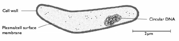
The diagram shows the bacterium Mycobacterium tuberculosis, which causes tuberculosis (TB).
(a) M. tuberculosis is taken up by macrophages and multiplies inside them.
After 4 hours, the macrophages were sampled to find out how many had taken up either glass beads or bacteria. The results are shown in the graph. The x-axis shows the initial ratio of bacteria or glass beads to macrophages in the mixture.
Discuss what these results suggest about the ability of macrophages to take up M. tuberculosis.
(c) When M tuberculosis is present inside a phagosome of a macrophage, it secretes glycolipids that accumulate in lysosomes and prevent the lysosomes fusing with the phagosome.
Explain how this prevents the macrophage from destroying the bacterium. (3 marks)
Candidate A
(a) It stops the B cells seeing them, so they don't make antibodies ü against them.
* This is not a very clear answer. B cells do not 'see', so this is not a good term to use. The 'they' in the second half of the sentence could refer to either B cells or the bacteria. 1/3
(b) The macrophages took up more glass beads than bacteria .ü So they are not very good at taking up the bacteria .ü
* Just enough for two marks, although the second sentence is weak. 2/3
(c) tysosornes contain digestive enzymes, ü so if they don't fuse with the phagosome the bacteria won't get digested. ü
* Once again, the candidate has the right ideas, but does not give enough biological detail to get full marks. 2/3
Candidate B
(a) B cells only become active when they meet the specific antigen ü to which they are able to respond. If the bacteria are inside a macrophage. then the B cell's receptors won't meet the antigen ü on the bacteria. This means that the B cells will not divide to produce plasma cells , üand will not secrete antibodies üagainst the bacteria.
* A good answer. 3/3
(b) The cells only started to take up any bacteria when the particle.macrophage ratio was 1 ü On the other hand, they took up glass beads even when the ratio was above 0.01. üWhen the ratio of particles to macrophages was 10, only about 30% of the macrophages had taken up bacteria, whereas over 75% of them had taken up glass beads. ü This shows the macrophages do take up the bacteria,
but not as well as they take up glass beads. ü
* A good answer, which does attempt to 'discuss' by providing statements relating to the relatively low ability of the macro phages to take up the bacteria, but also stating that they do take them up. In general, it is always a good idea to quote data where they are relevant in your answer. 3/3
(c) Normally, lysosomes fuse with phagosomes and release hydrolytic enzymes üinto them. These enzymes then hydrolyse (digest) whatever is in the phagosome. ü If this doesn't happen, then the bacteria can live inside the phagesome ü without being digested.
* All correct. 3/3
(a) M. tuberculosis is taken up by macrophages and multiplies inside them.
Suggest how this strategy could help to protect M. tuberculosis from the immune response by B cells. (3 marks)
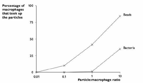
(b) In an experiment to investigate how M tuberculosis avoids destruction by macrophages, bacteria were added to a culture of macrophages obtained from the alveoli of mice. At the same time, a quantity of small glass beads, equivalent in size to the bacteria, were added to the culture. The experiment was repeated using increasing quantities of bacteria and glass beads.After 4 hours, the macrophages were sampled to find out how many had taken up either glass beads or bacteria. The results are shown in the graph. The x-axis shows the initial ratio of bacteria or glass beads to macrophages in the mixture.
Discuss what these results suggest about the ability of macrophages to take up M. tuberculosis.
(3 marks)
(c) When M tuberculosis is present inside a phagosome of a macrophage, it secretes glycolipids that accumulate in lysosomes and prevent the lysosomes fusing with the phagosome.
Explain how this prevents the macrophage from destroying the bacterium. (3 marks)
Total: 9 marks
Candidate A
(a) It stops the B cells seeing them, so they don't make antibodies ü against them.
* This is not a very clear answer. B cells do not 'see', so this is not a good term to use. The 'they' in the second half of the sentence could refer to either B cells or the bacteria. 1/3
(b) The macrophages took up more glass beads than bacteria .ü So they are not very good at taking up the bacteria .ü
* Just enough for two marks, although the second sentence is weak. 2/3
(c) tysosornes contain digestive enzymes, ü so if they don't fuse with the phagosome the bacteria won't get digested. ü
* Once again, the candidate has the right ideas, but does not give enough biological detail to get full marks. 2/3
Candidate B
(a) B cells only become active when they meet the specific antigen ü to which they are able to respond. If the bacteria are inside a macrophage. then the B cell's receptors won't meet the antigen ü on the bacteria. This means that the B cells will not divide to produce plasma cells , üand will not secrete antibodies üagainst the bacteria.
* A good answer. 3/3
(b) The cells only started to take up any bacteria when the particle.macrophage ratio was 1 ü On the other hand, they took up glass beads even when the ratio was above 0.01. üWhen the ratio of particles to macrophages was 10, only about 30% of the macrophages had taken up bacteria, whereas over 75% of them had taken up glass beads. ü This shows the macrophages do take up the bacteria,
but not as well as they take up glass beads. ü
* A good answer, which does attempt to 'discuss' by providing statements relating to the relatively low ability of the macro phages to take up the bacteria, but also stating that they do take them up. In general, it is always a good idea to quote data where they are relevant in your answer. 3/3
(c) Normally, lysosomes fuse with phagosomes and release hydrolytic enzymes üinto them. These enzymes then hydrolyse (digest) whatever is in the phagosome. ü If this doesn't happen, then the bacteria can live inside the phagesome ü without being digested.
* All correct. 3/3
- #84 Question 7
Beetroot cells contain a red pigment that cannot normally escape from the cells through the cell surface membrane. A student carried out an investigation into the effect of temperature on the permeability of the cell surface membrane of beetroot cells....
- #82 Question 5
The diagram below shows a small part of a human lung as it appears through a microscope. (a) Name the type of blood vessel in which the red blood cell is present. (1 mark)(b) Describe and explain two ways in which the structure of the alveoli, shown...
- #80 Question 3
(a) The diagrams show a cell in various stages ofthe mitotic cell cycle. Name the stage represented by each diagram, and arrange them in the correct sequence. (b) Describe the role of spindle microtubules in mitosis. (3 marks) (c) The graph below shows...
- #78 Question 1
(a) The diagram shows a small part of a cell, as seen using an electron microscope. (i) Name the parts labelled A to D. (2 marks) (ii) Describe how part B is involved in the formation of extracellular...
- Prokaryote: Cells
In this picture are millions and millions of bacteria. No matter how much I clean the countertops, once an object or hand is placed on it, more bacteria live there. Bacteria is a prokaryote which are cells that do not have a nucleus or membrane bound...