Biology
 The kidneys remove wastes from the blood and are the effectors for controlling the water potential of the blood. The removal of waste products generated by metabolic reactions inside body cells is called Excretion. Some of these products are toxic, while others are simply in excess of requirements.
In mammals, the 2 major excretory products are:
The structure and histology of kidneys
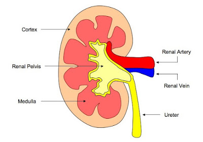
Each kidney is supplied with oxygenated blood through a renal artery. Blood is removed in the renal vein. A tube called the ureter takes urine from the kidney to the bladder.
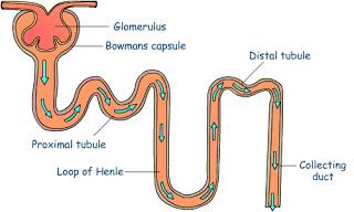
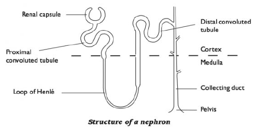
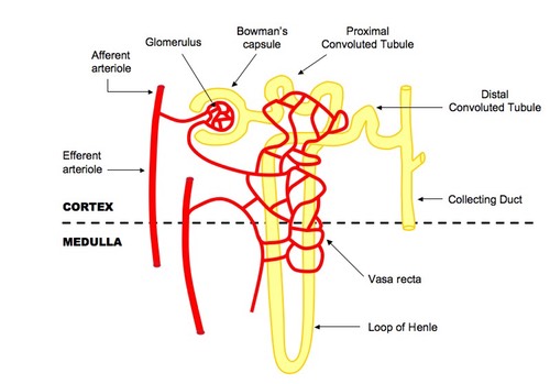
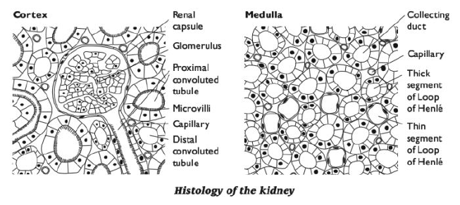
Each kidney contains thousands of microscopic tubes called nephrons. The beginning of each nephron is a cup-shaped structure called a renal capsule (Bowman's capsule). This is in the cortex of the kidney. The tube leads from the renal capsule down into the kidney medulla, then loops back into the cortex before finally running back down through the medulla into the pelvis of the kidney, where it joins the ureter.
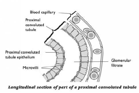
Each nephron has a network of blood vessels assodated with it. Blood arrives in the afferent arteriole (from the renal artery), and is delivered to a network of capillaries, called a glomerulus, in the cup of the renal capsule. Blood leaves the glomerulus in the efferent arteriole, which is narrower than the afferent arteriole. This leads to another network of capillaries that wraps around the nephron, before delivering the blood to a branch of the renal vein.
The diagrams below show the histology (structure of tissues) of the kidney.
- #113 The Control Of Blood Glucose
The blood glucose concentration is regulated by negative feedback control mechanisms. Blood glucose concentration should remain at a fairly constant value of about 100 mg glucose per 100 cm3 of blood. If blood glucose concentration falls well below...
- #112 Osmoregulation
Osmoregulation is the control of the water content of body fluids. It is part of homeostasis, the maintenance of a constant internal environment. It is important that cells are surrounded by tissue fluid of a similar water potential to their own...
- #111 Production Of Urine In A Nephron - Ultrafiltration And Reabsorption
Ultrafiltration occurs at the barrier between the blood and the filtrate in the renal capsule or Bowman's capsule in the kidneys. Ultrafiltration The Bowman's capsule contains a dense capillary network called the glomerulus. Blood...
- #109 Thermoregulation - The Control Of Body Temperature
One of the most important examples of homeostasis is the regulation of body temperature. It involves both coordination systems - nervous and endocrine. Not all animals can do this physiologically. Endotherms - Animals (e.g. birds and mammals)...
- #108 Homeostasis In Mammals
In the body of an animal conditions such as water concentration, temperature, and glucose concentration must be kept as constant as possible. Control systems that keep such conditions constant are examples of homeostasis; this is the maintenance of constant...
Biology
#110 Excretion and structure of Kidneys
In mammals, the 2 major excretory products are:
- CO2 produced by aerobic respiration. CO2 dissolves in H2O to produce a weak acid, so if too much builds up in body fluids the pH drops, which can damage cells and disrupt metabolism. CO2 is transported to the lungs dissolved in blood plasma and excreted in expired air.
- Nitrogenous excretory products, in particular urea. Excess amino acids cannot be stored in the body. In the liver, they are converted to urea, CO(NH2)2, and a keto acid. The keto acid can be respired to provide energy, or converted to fat for storage. The urea dissolves in the blood plasma and is removed and excreted by the kidneys.
The structure and histology of kidneys
Each kidney is supplied with oxygenated blood through a renal artery. Blood is removed in the renal vein. A tube called the ureter takes urine from the kidney to the bladder.
The diagrams below show the histology (structure of tissues) of the kidney.
Homeostasis in mammals requires complex systems to maintain internal conditions near constant. The kidneys remove wastes from the blood and are the effectors for controlling the water potential of the blood. a) discuss the importance of homeostasis in mammals and explain the principles of homeostasis in terms of internal and external stimuli, receptors, central control, co-ordination systems, effectors (muscles and glands) b) define the term negative feedback and explain how it is involved in homeostatic mechanisms c) outline the roles of the nervous system and endocrine system in co-ordinating homeostatic mechanisms, including thermoregulation, osmoregulation and the control of blood glucose concentration d) describe the deamination of amino acids and outline the formation of urea in the urea cycle (biochemical detail of the urea cycle is not required) e) describe the gross structure of the kidney and the detailed structure of the nephron with its associated blood vessels using photomicrographs and electron micrographs f) describe how the processes of ultrafiltration and selective reabsorption are involved with the formation of urine in the nephron g) describe the roles of the hypothalamus, posterior pituitary, ADH and collecting ducts in osmoregulation h) explain how the blood glucose concentration is regulated by negative feedback control mechanisms, with reference to insulin and glucagon i) outline the role of cyclic AMP as a second messenger with reference to the stimulation of liver cells by adrenaline and glucagon j) describe the three main stages of cell signalling in the control of blood glucose by adrenaline as follows: ? hormone-receptor interaction at the cell surface ? formation of cyclic AMP which binds to kinase proteins ? an enzyme cascade involving activation of enzymes by phosphorylation to amplify the signal k) explain the principles of operation of dip sticks containing glucose oxidase and peroxidase enzymes, and biosensors that can be used for quantitative measurements of glucose in blood and urine l) explain how urine analysis is used in diagnosis with reference to glucose, protein and ketones |
- #113 The Control Of Blood Glucose
The blood glucose concentration is regulated by negative feedback control mechanisms. Blood glucose concentration should remain at a fairly constant value of about 100 mg glucose per 100 cm3 of blood. If blood glucose concentration falls well below...
- #112 Osmoregulation
Osmoregulation is the control of the water content of body fluids. It is part of homeostasis, the maintenance of a constant internal environment. It is important that cells are surrounded by tissue fluid of a similar water potential to their own...
- #111 Production Of Urine In A Nephron - Ultrafiltration And Reabsorption
Ultrafiltration occurs at the barrier between the blood and the filtrate in the renal capsule or Bowman's capsule in the kidneys. Ultrafiltration The Bowman's capsule contains a dense capillary network called the glomerulus. Blood...
- #109 Thermoregulation - The Control Of Body Temperature
One of the most important examples of homeostasis is the regulation of body temperature. It involves both coordination systems - nervous and endocrine. Not all animals can do this physiologically. Endotherms - Animals (e.g. birds and mammals)...
- #108 Homeostasis In Mammals
In the body of an animal conditions such as water concentration, temperature, and glucose concentration must be kept as constant as possible. Control systems that keep such conditions constant are examples of homeostasis; this is the maintenance of constant...