Biology
- Prokaryotes Vs Eukaryotes
While eukaryotic cells and prokaryotic cells have some characteristics in common, they diverged from their common ancestor billions of years ago, thus accounting for significant differences in overall structure and function. Here we will go over them....
- Animal Eukaryotic Cell Components: Structure And Function
I posted a picture of the eukaryotic animal cell earlier, but it was simply a labeled diagram. Here we will go into each of the major parts of the cell, as well as each organelle's function, not to mention the overall structure of the cell. As the...
- Eukaryotic Cells
Cell Webquest ? Ms. Carter Eukaryotic Cells California Standard: Cell Biology 1.a, e, f, g CELL BIOLOGY 1 a. The fundamental life processes of plants and animals depend on a variety of chemical reactions that occur in specialized areas of the organism?s...
- Skin And Hair Facts
A lifespan of an eyelash is approximately 150 days.A Russian man who wore a beard during the time of Peter the Great had to pay a special tax.A survey done by Clairol 10 years ago came up with 46% of men stating that it was okay to color their hair. Now...
- When Is A Chloroplast Not A Chloroplast?
Biology concepts ? gravitropism, plastid, chloroplast, chromoplast, amyloplast, leucoplast, malaria parasite Believe it or not, the way plant roots know to grow into the dirt is related to photosynthesis! ?How can this be?? you ask. Well, let?s talk about...
Biology
Cell To Cell Tanning
Biology Concepts ? vacuoles, phagocytosis, melanin, cephalopod camouflage
 |
Sometimes, the name of an item becomes the same as its function. Have you ever asked for a Puff when you needed a facial tissue? But we all ask for a Kleenex. Vacuoles are the same, but in reverse, they are all very similar, but are named based on their function. |
Last week we discussed the specific characteristics of organelles that allow them and the cells in which they work to specialize their activities. But being specific is not always more accurate. We ask for a Kleenex no matter what brand of facial tissue is handy. Many people ask for a Coke, when they really mean any soda at all.
There is an organelle that gets the same treatment. The vacuole is a generic membrane bound compartment; its function and name brand is usually defined by what it is carrying. Lysosomes are vacuoles, named for the lysosomal enzymes they carry. Peroxisomes are vacuoles, named for the peroxidase enzymes they carry. Branding can be important, even in biology.
 |
The vacuole is a membrane bound compartment inside the cell. The central vacuole of the plant cell is shown above, and can occupy a huge percentage of the cell volume. It can store water or carbohydrates, and can change size very rapidly. |
Most cell types have many vacuoles or vesicles (small vacuoles), but there are exceptions. Plant cells generally just have one vacuole - not one type - but a total number of 1. This is called the central vacuole, and can occupy up to 90% of the cell volume. We have discussed its functions several times; the central vacuole is the part of the cell that fills and empties in order to induce movement of plant components, like flowers blooming and leaves folding (Plants That Don?t Sleep). Vacuole functions seem to be quite varied, from digestion to structure.
The melanosome is another type of vacuole. Take a guess how it got its name?? O.K., it?s because it carries melanin. Melanin (melas in Greek means black) is the pigment that gives color to your skin, hair, iris (the colored part of your eye), and even parts of your brain. One crucial function of melanin is to protect your skin cells from ultraviolet (UV) damage. This is why natives in sunnier areas have darker skin, while people from latitudes farther north and south have lighter skin tones.
 |
Inuit (translated as ?the people?) are a group of different peoples of the north latitudes. In Alaska, the term Eskimo is often used because its meaning includes the two main groups that live there. All Inuit seem to have darker skin tones than one would think was called for. |
However, even in this there is an exception. Inuit natives in the Arctic have darker skin on average as compared to white Americans or Europeans. This may be because the amount of sunlight that reflects off the snow increases their UV exposure, or because they have been living in this environment for only 5000 years and haven?t had time to adopt a lighter skin color. Either way, you don?t see many red-headed, freckled, Inuits.
In your skin, melanin is produced by a specialized skin cell type called the melanocyte. We discussed in last week?s post that the different combinations of organelles allows cells to take on specialized function, and this is the case with melanocytes. Melanin is synthesized from the amino acid tyrosine within the melanosome, and remains stored in this organelle when it is stored, moved, and when it performs it jobs.
Only 5-10% of skin cells are melanocytes, and they are located only in the deepest layer of skin, called the stratum basale. (stratum = layer and basale = base or lowest) But if you look at dark skin or at a well-tanned person, it seems that there is pigment all over. You would think that freckles would be more natural, the melanocytes that produce melanin are spotted over the skin, and so are freckles. So how does a person get tanned skin evenly, or how is it that dark complexions are homogeneous over a large area?
This is where the melanosomes as organelles are so exceptional; they can move from melanocytes to keratinocytes (skin cells). This is unique amongst organelles and is still not fully understood. However, much evidence has been uncovered in just the past few years.
 |
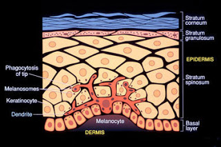
Melanocytes are rare in the skin, but can project up into the upper layers of the skin in order to spread out their melanin. |
The melanocyte is stimulated to make melanin due to UV exposure of keratinocytes. When the DNA of the skin cell is hit with UV radiation, it triggers production of a hormone called alpha-melanocyte stimulating hormone, alpha-MSH. This hormone acts on the melanocytes through receptors on the cell membrane (2nd messenger system, see Multitaskers). The message is transferred to the melanocyte nucleus and melanin is produced in melanosomes.
Next, the melanosome grows dendrites (from Greek for tree), sometimes called filipodia (like a foot). These extensions snake their way between keratinocytes and reach up into the higher layers of skin cells. The dendrites are rich in melanosomes, but research has yet to show if the melanosomes are responsible for the formation of the dendrites. This may be so, because it is not until the keratinocytes acquire melanosomes that they also start to form these filopodia.
The melanosomes end up inside the skin cells, and this is where current research is focused. Recent hypotheses for their movement include ideas that they are released from melanocytes and then taken up by keratinocytes, or that there is fusion of the keratinocyte and the melanocyte.
However, evidence from a 2010 study indicates that the keratinocyte actually swallows (phagocytoses, phag = eat and cyto = cell) the ends of the dendrites, and the included melanosomes become skin cell organelles. The keratinocyte membrane expands around the end of the dendrite, then pinches together until the two sides meet each other. Part of the melanocyte, its cytoplasm, its membrane, and its melanosomes ends up as part of the keratinocyte. Your trip to the beach causes your cells to eat each other, cool!
The phagocytosed melanosomes can have two fates, but neither is what you would expect. Usually phagocytosed vacuoles are merged with lysosomes and the contents are degraded. But not so for the melanosome.
Some are pushed out into new keratinocyte filopodia. These dendrites can then be phagocytosed by other keratinocytes. In this way, the melanin produced by the few melanocytes can be spread through out the layers and surface area of the skin cells and result in a continuous skin tone. However, if you have fewer melanocytes, or they have a mutated alpha-MSH receptor that forces them to produce more local melanin, you end up with freckles.
 |
These micrographs show that melanosomes aggregate around the nucleus of keratinocytes that have been exposed to UV radiation in order to protect the DNA in the nucleus. |
The second fate for the melanosomes occurs when they get the right signal, and is just as amazing. The UV rays that can stimulate the DNA to make alpha-MSH can also induce DNA damage; this is the main reason for melanin production. In response to increased sun exposure, the keratinocytes that take up the melanosomes will move them into position between the UV source and their DNA, like a hat worn by the nucleus. There the melanin absorbs the radiation like nature?s own sunscreen.
In truth, melanin is really three different pigments. Eumelanin (eu = true) is dark brown and is the most common type of melanin. But the same cells and melanosomes also produce pheomelanin (pheo = dusky) which is more reddish in color. Pheomelanin is responsible for red hair and for the freckle color in fair-skinned individuals.
Finally, there is neuromelanin in the brain, which gives a dark color to the portions of the brain like the substantia nigra (Latin for black substance). This brain structure coordinates muscle movement, and when these cells die or malfunction, the result is Parkinson?s disease. The melanin in these cells is actually just a byproduct of dopamine production. Parkinson?s disease can be treated, at least in its early stages, with synthetic dopamine.
Although it serves no known function in the midbrain, melanin does help in ways other than UV absorption. Different stressors, like chemicals, oxidative damage, and high temperature are also suppressed by melanin.
Melanin is particularly important for cephalopods, like squid, cuttlefish, and octopuses (yes, the plural of octopus is octopuses or octopodes, not octopi). These animals can disguise themselves within their environments and this task requires melanin. Under their skin, cephalopods have three or four layers of cells that allow them to create many colors and surface characteristics.
Chromatophores are the top level. The have saccules of melanin that can change shape. When stretched out, they show much color, but when relaxed, only pinpoints of color show. Each saccule is attached to many muscles and each muscle is innervated by several neurons; the octopus has fine control of each and every saccule, and each saccule can rapidly assume a different size and shape. This makes many shades and patterns possible
 |
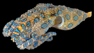
Cephalopods use several specialized cell types to hide themselves from predators or to display for a mate or rival. These special functions are possible, in part, because of the organelles they possess. In the case of the blue ring octopus, the blue is not from melanin, and is used to warn of its toxicity, not to hide. |
Below the chromatophores are the iridophores. These are like little mirrors that can also change angle position through many muscle and nerve controls. This allows the cephalopod to change the reflectivity and shine of its surface. Under this layer are the leucophores. They specialize in reflecting the dominant light wavelengths that they receive. These are the cells that help the octopus to match its surrounding colors so well (amazing video). Lastly, only some cephalopods have a layer of photophores that produce light (bioluminescence). We will talk about this amazing feat in a future series of posts.
Working together, and coordinated by their fantastic eyes, cephalopod skin cell layers can perform some amazing tricks to help the organism to survive. All this muscle control and sensory input requires a big brain, and cephalopods have the biggest brains of all the invertebrates (no backbone) and bigger than many vertebrates. I think they should be worthy of a post or two in the future, especially since almost all cephalopods are color blind!
But back to melanin. Squid and octopus ink is also made of melanin, and is most often used to confuse predators, but I especially like its effect when used with eggs, water, and flour ? try squid ink pasta sometime, you?ll like it.
Finally, there is a new way that melanin is helping science. Melanosomes are big, and they tend to fossilize well, so scientists are starting to learn what the coloration of dinosaurs might have been based on the preserved melanosomes and their included melanins.
We have looked at several organelle types, and have seen how they allow for specialized cell functions. But what if you had to get along without organelles, could a cell cope? We'll see about this next week.
Suman K. Singh, Robin Kurfurst, Carine Nizard, Sylvianne Schnebert, Eric Perrier and Desmond J. Tobin (2010). Melanin transfer in human skin cells is mediated by filopodia?a model for homotypic and heterotypic lysosome-related organelle transfer FASEB Journal DOI: 10.1096/fj.10-159046
For more information and classroom activities on vacuoles, melanin, melanosome movement, or cephalopod camouflage, see:
Vacuoles ?
http://www.biology4kids.com/files/cell_vacuole.html
http://kentsimmons.uwinnipeg.ca/cm1504/vacuoles.htm
http://micro.magnet.fsu.edu/cells/plants/vacuole.html
http://www.nature.com/scitable/topicpage/plant-vacuoles-and-the-regulation-of-stomatal-14163334
http://bcs.whfreeman.com/thelifewire/content/chp28/2802001.html
http://www.bscb.org/?url=softcell/vacuole
http://library.thinkquest.org/27819/ch3_3.shtml
http://www.illuminatedcell.com/Vacuolarsystem.html
http://cfeil.tripod.com/vacuoles.htm
Melanin ?
http://www.pbs.org/race/000_About/002_04-teachers-06.htm
http://www.healthofchildren.com/A/Albinism.html
http://www.webexhibits.org/causesofcolor/7F.html
http://www.buzzle.com/articles/enzymatic-browning.html
http://www.google.com/url?sa=t&rct=j&q=&esrc=s&source=web&cd=28&ved=0CHIQFjAHOBQ&url=http%3A%2F%2Fwww.curriculumsupport.education.nsw.gov.au%2Fsecondary%2Fscience%2Fassets%2Faifst%2FExperiments%2Fapple_browning.pdf&ei=du5lT_zTHMXdtgen34z-DQ&usg=AFQjCNHdPQWxis8p_rvs8t0bWgNg3yqyWQ
http://www.cl.cam.ac.uk/~jgd1000/melanin.html
http://www.sciencedaily.com/releases/2010/10/101014171144.htm
http://www.clinuvel.com/en/dermatology/melanin/function-skin-pigment
http://www.rastafarian.net/what_is_melanin.htm
Melanosome movement ?
http://jcs.biologists.org/content/121/24/3995.full
http://news.nationalgeographic.com/news/2010/01/100127-dinosaur-feathers-colors-nature/
http://en.wikipedia.org/wiki/Griscelli_syndrome
http://www.medscape.com/viewarticle/462276_3
Cephalopod camouflage ?
www.thecephalopodpage.org/.../HowCephalopodsChangeColor.pdf
www.thecephalopodpage.org/cephschool/CephalopodVision.pdf
www.thecephalopodpage.org/.../ColorChangeInCephalopods.pdf
http://nationalzoo.si.edu/Animals/Invertebrates/Facts/cephalopods/colordisguise.cfm
http://www.mbl.edu/mrc/hanlon/coloration.html
http://www.talkingscience.org/2011/01/are-cephalopods-really-colorblind/
http://marinediscovery.arizona.edu/lessonsF00/blennies/2.html
http://www.oercommons.org/courses/cephalopod-lesson-plans
http://tolweb.org/treehouses/?treehouse_id=4225- Prokaryotes Vs Eukaryotes
While eukaryotic cells and prokaryotic cells have some characteristics in common, they diverged from their common ancestor billions of years ago, thus accounting for significant differences in overall structure and function. Here we will go over them....
- Animal Eukaryotic Cell Components: Structure And Function
I posted a picture of the eukaryotic animal cell earlier, but it was simply a labeled diagram. Here we will go into each of the major parts of the cell, as well as each organelle's function, not to mention the overall structure of the cell. As the...
- Eukaryotic Cells
Cell Webquest ? Ms. Carter Eukaryotic Cells California Standard: Cell Biology 1.a, e, f, g CELL BIOLOGY 1 a. The fundamental life processes of plants and animals depend on a variety of chemical reactions that occur in specialized areas of the organism?s...
- Skin And Hair Facts
A lifespan of an eyelash is approximately 150 days.A Russian man who wore a beard during the time of Peter the Great had to pay a special tax.A survey done by Clairol 10 years ago came up with 46% of men stating that it was okay to color their hair. Now...
- When Is A Chloroplast Not A Chloroplast?
Biology concepts ? gravitropism, plastid, chloroplast, chromoplast, amyloplast, leucoplast, malaria parasite Believe it or not, the way plant roots know to grow into the dirt is related to photosynthesis! ?How can this be?? you ask. Well, let?s talk about...
