Biology
The differences between the various prion diseases are based on the specific prion protein mutation, what part of the brain is attacked, and how potent the prion is at refolding normal prion proteins. For instance, the D178N mutation in FFI also occurs in Creutzfeldt-Jakob Disease (CJD), but a normal polymorphism (an amino acid change that doesn?t change form or function) at position 129 determines the fate. If amino acid 129 is methionine, the the person gets FFI, if it is valine, then they get CJD.
The families that suffer from FFI have the D178N mutation, and also pass on the polymorphism for methionine (M) at position 129. Even more gruesome, some cases of prion protein diseases can be sporadic, not associated with either an inherited mutation or transmission. The malfolded prion can very rarely arise out of nowhere in isolated individuals.
On autopsy, the hypothalmus of an FFI sufferer looks like it has been hit with a shotgun blast. Holes are present in the tissue, representing areas where neurons have been lost due to inflammation and triggered cell death. The affected area of the brain takes on a spongy appearance, so prion protein diseases are lumped together and called transmissable spongiform encephalopathies (encephalon = brain and pathy = disease). Unfortunately, there are no cure, treatments, or vaccines for any of these prion diseases.
- Gene Mutation
A gene mutation is a change in one or several bases. A base may be added, deleted, or substituted with another base. This is caused often through the action of damaging chemicals, radiations, or through errors inherent in DNA replication and repair reactions....
- Three Codons Terminate Protein Synthesis
KEY TERMS:The amber codon is the triplet UAG, one of the three termination codons that end protein synthesis. The ochre codon is the triplet UAA, one of the three termination codons that end protein synthesis. The opal codon is the triplet UGA, one of...
- Mutations May Cause Loss-of-function Or Gain-of-function
KEY TERMS:A null mutation completely eliminates the function of a gene. Leaky mutations leave some residual function, for instance when the mutant protein is partially active (in the case of a missense mutation), or when read-through produces a small...
- # 36 Gene Mutation, Sickle Cell Anaemia
A gene mutation is a change in the sequence of nucleotides that may result in an altered polypeptide. A mutation is a random, unpredictable change in the DNA in a cell. It may be: ? a change in the sequence of bases in one part of a DNA molecule?...
- Proteins
PROTEINSProteins are polypeptides. i.e., linear chains of amino acids linked by peptide bonds. A Peptide bond is formed when ?COOH group of one amino acid reacts with ?NH2 group of next amino acid by releasing a molecule of water (dehydration). Proteins...
Biology
An Infectious, Genetic Disease? Better Sleep On It.
Biology concepts ? thermoregulation, sleep, genetic disease, infectious disease, central dogma of molecular biology, form follows function
 |
Even rats have to get some sleep. It was nice to have the sleeping cap, but unnecessary for a sleep deprivation study. Not a good use of research dollars. |
?I?m dying for a good night?s sleep.? Is this just hyperbole, or an impending warning of death? For laboratory rats, sleep deprivation does kill. During their insomniac downward spiral, the rats tend to get hot and can?t cool down ? you know, they can't thermoregulate (see Can?t We Just Go With The Flow). This doesn?t mean that a loss of the ability to thermoregulate is what kills the rats, but it does suggest a connection between sleep deprivation and the hypothalamus.
We looked at the hypothalamus in our story of endothermy. This evolutionarily old brain structure implements a set point temperature for the body and receives information about the temperature of different parts of the body. When the body temperature deviates from the set point, the hypothalamus initiates bodily mechanisms to normalize the temperature.
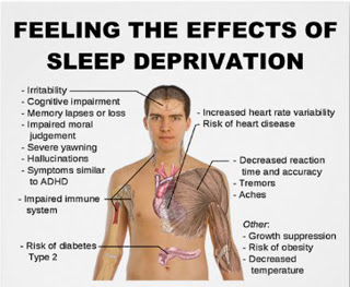 |
Apparently one of the effects of sleep deprivation is that you become semi-transparent. |
People with severe insomnia tend to sweat more and have higher core temperatures even though they say they are cold. They also have extreme high blood pressure, pulse, and appetite. These symptoms suggest that sleep deprivation messes with the hypothalamus, since functions of the hypothalamus include themoregulation, sleep, hunger, thirst, reproductive readiness in females, and stress responses. What scientists don?t know yet is just how sleep deprivation actually kills the rats or harms people.
Dying from a lack of sleep is not just a rat problem, a few very unlucky humans die from it as well. Fatal familial insomnia (FFI) is a very rare genetic disorder; it has been reported in only 40 families worldwide. Before describing the truly horrible way these patients die, let?s look at what causes the disease.
FFI is caused by a point mutation in the gene for the prion protein PrPc. A point mutation means that one nucleotide on the DNA is changed, which leads to a change in the protein coded for by the DNA. Three unit (nucleotides) segments of the RNA (made from the DNA template) work together (called a codon) to code for one protein building block (amino acid). In the case of FFI, the amino acid called aspartic acid is changed to one called asparagine, and this changes the protein?s shape.
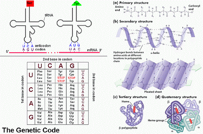 |
The left image shows mRNA bases recognized in sets of three (codons) by tRNAs with amino acids attached (Ser = serine, tyr = tyrosine). The amino acids are linked to because proteins. The lower section is the genetic code, showing which amino acids are coded for by which codons. The right image shows how proteins fold. The primary structure is the amino acid sequence. The secondary structure comes from interactions of adjacent amino acids, including spirals called helices or sheets. The tertiary structure comes from the folding up of the entire protein, while the quaternary structure comes from the interaction of different proteins into a larger complex. |
PrPc is made up of 250 amino acids linked together in a chain. Each different amino acid carries a different shape and charge and will interact with every other amino acid differently. The sequence of amino acids in a protein cause it to fold into a specific shape. It is the protein?s conformation (shape) that determines its function. This is the opposite of what we determined for evolved organism characteristics, where form follows function (see Do You Have To Be Ugly To Hear Well?). With proteins ? function follows form!
Mutation of that single amino acid at position 178 (aspartic acid is negatively charged, while asparagine is positive) causes the folding, and therefore the function, of the protein to change. Aspartic acid is sometimes abbreviated "D", while asparagine is called "N"; therefore, the mutation is often indicated as D178N (D at position 178 is changed to N).
Many genetic mutations result in no change in amino acid, or a change that bring a large enough change the shape to cause a change in function. But when it does, good or bad things can happen. On one hand, the altered protein might confer an advantage to the organism, one that promotes survival in the environment or after an environmental change.This positive selection through reproductive advantage become the new normal ? and this is evolution.
On the other hand, the change in amino acid sequence, form, and function could be destructive. Disease might be the result, or perhaps a change in the organism that reduces reproductive success. One of these two results is what occurs with the FFI mutation of the prion protein.
On the other hand, the change in amino acid sequence, form, and function could be destructive. Disease might be the result, or perhaps a change in the organism that reduces reproductive success. One of these two results is what occurs with the FFI mutation of the prion protein.
When the mutated prion folds differently, it forgets its day job and moonlights as a sinister evil force. Every other prion protein it contacts, WHETHER MUTATED OR NOT, is coaxed into changing its shape. The new prions turn to the dark side, then change other prion proteins they contact, multiplying the effect. The poorly folded prion proteins will stick together, come out of solution, and form solids (plaques) where they settle out. In different prion protein diseases, this settling out occurs in different parts of the brain. In FFI, it is the hypothalamus.
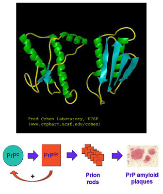 |
In the top image, the PrPc on the left is properly folded. The green represents alpha helices and the blue arrows represent beta-pleated sheets. The right image shows the malfolded version of PrPsc. It is a tighter structure, which partially explains why protein-degrading enzymes don?t work on it. . The lower cartoon shows that the PrPsc can force the PrPc to assume the improper form, and these then aggregate into plaques. |
The prion plaques are longer lived then the regular prion protein; normal cellular enzymes whose job it is to degrade proteins won?t work on prion plaques. And worse, if some of the malfolded protein is transferred to another animal, the recipient will develop plaques and disease as well. That makes this an infectious disease that isn?t caused by a bacteria, fungus, parasite, or virus. The prion is an infectious protein! What a terrible exception to the rules of infectious diseases.
We see here a protein that can replicate itself (not by building more of themselves, but by changing the form of normal proteins), and that makes it a repository of biologic information. This is an exception to the central dogma of molecular biology, which says that DNA is the sole information storing material.
FFI moves from person to person through heredity, but if a non-affected person comes into contact with some brain material from an FFI patient and that material entered their bloodstream, it can be transmitted this way as well. A prion protein disease called Kuru is famous for being transmitted from person to person.
The Fore tribe in Papua New Guinea once observed a ritual wherein they honored a dead tribe member by eating part of their brain (called ritualistic mortuary cannibalism - gasp!). Because of this, there was an epidemic of Kuru in this tribe in the early 1900?s. Over a period of 3-6 months victims would become unsteady, irrational with bouts of laughter, and then degrade mentally and physically to the point of death. There are more than twenty known prion diseases (mad cow disease, Creutzfeldt-Jakob, scrapie, etc.), and Kuru suggests that some might have no genetic component, only person to person transmission.
 |
A member of the Fore tribe is shown on the left. This tribe used to celebrate the lives of departed members by eating their brains. This spread a prion protein disease called Kuru, a protein disease that is infectious! The Fore tribe still lives in Papua New Guinea, although there are fewer of them than before Kuru. |
The families that suffer from FFI have the D178N mutation, and also pass on the polymorphism for methionine (M) at position 129. Even more gruesome, some cases of prion protein diseases can be sporadic, not associated with either an inherited mutation or transmission. The malfolded prion can very rarely arise out of nowhere in isolated individuals.
The mutated PrPc is passed on via inheritance. You get one copy of each chromosome from each of your parents, so for an individual gene, you might get two normal copies, 1 mutant copy and 1 normal copy, or 2 mutant copies. Some diseases require that you must inherit two mutant copies for symptoms to show (recessive), but other require only one mutant copy (dominant, it dominates the trait from the other parent).
FFI is autosomal dominant (not associated with the X or Y sex chromosomes), so the chance of getting a mutant copy and the disease if one parent has it is 1 in 2; these are bad odds. But, if everyone with FFI dies, then why is the disease still showing up in families. Remember that we said above that some genetic diseases can, but don't have to, affect reproductive success. Unfortunately for those with FFI, the symptoms appear in the victims? fifties, after they have had children. Natural selection doesn?t eliminate FFI from the population because FFI doesn?t appear affect reproduction.
The first symptoms of FFI include sweating while feeling cold. Later, the ability to get a good night?s sleep is lost, followed closely by the inability to nap. As the disease progresses, there are panic attacks, phobias, and no sleep whatsoever. After 4-6 months, mental abilities start to degrade. In its final stages unresponsiveness precedes death.
This is especially sad way to die, because during the majority of the disease course the patient is aware of everything going on. At least with middle to late Alzheimer?s disease the patient is blissfully unaware of their dementia.
This is especially sad way to die, because during the majority of the disease course the patient is aware of everything going on. At least with middle to late Alzheimer?s disease the patient is blissfully unaware of their dementia.
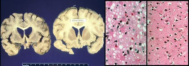 |
For both the gross and microscopic images, the left example is from prion protein disease victim, while the right example is from a normal brain. The brains on the left show how great the loss of tissue can be in Creutzfeldt-Jakob disease. The microscopic image from the diseased brain shows the plaques and the resulting holes in the brain structure. The small gaps in the normal brain on the right are a result of shrinking of tissue after it was on the slide. |
It is the hypothalamus' control of sleep cycles and circadian rhythms that promotes survival in animals. But what about plants? They don?t have a hypothalamus. Can they suffer from loss of circadian activity? In a word ? yes! And this will be our starting point next time.
For more information or classroom activities on prion proteins, central dogma, infectious or genetic disease, the genetic code or protein structure, see:
Prion protein and diseases ?
http://science.education.nih.gov/home2.nsf/Educational+ResourcesResource+FormatsOnline+Resources+High+School/D07612181A4E785B85256CCD0064857B
http://www.mad-cow.org/misfolding_review.html
http://www.pbs.org/wgbh/nova/madcow/prions.html
http://users.rcn.com/jkimball.ma.ultranet/BiologyPages/P/Prions.html
http://www.prioncentre.ca/
http://depts.washington.edu/daglab/research/prion.html
http://memory.ucsf.edu/cjd/overview/intro/forms/single
https://www.msu.edu/~hinzalia/
http://www.scripps.edu/philanthropy/priondiseases.html
http://www.mad-cow.org/june98_sci.html
http://www.sciencedaily.com/releases/2008/06/080617111831.htm
http://www.nih.gov/news/pr/may2005/niaid-24.htm
http://www.popsci.com/science/article/2010-01/devoid-dna-infectious-prions-evolve-anyhow
http://www.medicalnewstoday.com/articles/195971.php
central dogma of molecular biology ?
http://homepage.mac.com/hjconklin/ConkWare/CD_index.html
http://employees.csbsju.edu/hjakubowski/classes/chem%20and%20society/cent_dogma/olcentdogma.html
http://www.accessexcellence.org/RC/VL/GG/central.php
http://cnx.org/content/m11415/latest/
http://www.youtube.com/watch?v=-ygpqVr7_xs
http://www.nature.com/news/2011/110519/full/news.2011.304.html
http://www.cbs.dtu.dk/staff/dave/DNA_CenDog.html
http://www.indiana.edu/~ensiweb/connections/genetics/dna.les.html
http://mvhigh.net/jnevis/central_dogma_activity.htm
www.wm.edu/act2online/projects/centraldogma/centraldogma.ppt
infectious disease ?
http://www.medicinenet.com/infectious_disease/focus.htm
http://www.mayoclinic.com/health/infectious-diseases/DS01145
http://www.koshland-science-museum.org/exhib_infectious/
http://pathmicro.med.sc.edu/book/infectious%20disease-sta.htm
http://www.pbs.org/wgbh/nova/teachers/activities/3318_02_nsn.html
http://www.seplessons.org/node/226
www.iseesystems.com/community/.../Infectious_Disease_Activity.pdf
http://www.windows2universe.org/teacher_resources/infectious_disease.html
http://science-education.nih.gov/supplements/nih1/diseases/guide/activity2-1.htm
genetic disease ?
http://www.ornl.gov/sci/techresources/Human_Genome/medicine/assist.shtml
http://www.buzzle.com/articles/genetic-diseases-list-disorders.html
http://learn.genetics.utah.edu/content/disorders/whataregd/
http://www.geneticalliance.org/diseases
http://kidshealth.org/teen/your_body/health_basics/genes_genetic_disorders.html
http://www.kumc.edu/gec/lessons.html
http://www.teachersdomain.org/resource/tdc02.sci.life.gen.lp_disorder/
www.dnai.org/teacherguide/pdf/ts_tour.pdf
http://www.scienceinschool.org/print/1050
http://www.pbs.org/wgbh/nova/teachers/activities/3413_genes.html
http://www.accessexcellence.org/AE/AEPC/WWC/1994/problem_solving.php
http://www.pbs.org/wgbh/nova/teachers/activities/0406_01_nsn.html
genetic code ?
http://employees.csbsju.edu/hjakubowski/classes/ch331/protstructure/olprotfold.html
http://users.rcn.com/jkimball.ma.ultranet/BiologyPages/C/Codons.html
http://www.ncbi.nlm.nih.gov/Taxonomy/Utils/wprintgc.cgi
http://www.brooklyn.cuny.edu/bc/ahp/BioInfo/GP/GeneticCode.html
http://collegebio.wordpress.com/2010/09/22/how-the-genetic-code-became-degenerate/
http://www.biologie.uni-hamburg.de/b-online/e21/21a.htm
http://www.genomebc.ca/education/outreach-programs/trips-in-2011-2012/classroom-activities/
www.greatscience.com/biomed_tech/pdfs/puzzle_of_life.pdf
protein structure ?
http://webhost.bridgew.edu/fgorga/proteins/default.htm
http://www.vivo.colostate.edu/hbooks/genetics/biotech/basics/prostruct.html
http://themedicalbiochemistrypage.org/protein-structure.html
http://lectures.molgen.mpg.de/ProteinStructure/index.html
http://staff.jccc.net/pdecell/biochemistry/protstruc.html
http://www.johnkyrk.com/aminoacid.html
http://www.neurogems.org/protSim/
http://molo.concord.org/database/activities/236.html
http://nature.ca/genome/05/051/0511/0511_m103_e.cfm
http://www.utsouthwestern.edu/education/programs/stars/resources/other-classroom-activities.html
http://www.scienceteacherprogram.org/biology/biolps.html
nwabr.org/sites/default/files/learn/bioinformatics/AdvL5.pdf
- Gene Mutation
A gene mutation is a change in one or several bases. A base may be added, deleted, or substituted with another base. This is caused often through the action of damaging chemicals, radiations, or through errors inherent in DNA replication and repair reactions....
- Three Codons Terminate Protein Synthesis
KEY TERMS:The amber codon is the triplet UAG, one of the three termination codons that end protein synthesis. The ochre codon is the triplet UAA, one of the three termination codons that end protein synthesis. The opal codon is the triplet UGA, one of...
- Mutations May Cause Loss-of-function Or Gain-of-function
KEY TERMS:A null mutation completely eliminates the function of a gene. Leaky mutations leave some residual function, for instance when the mutant protein is partially active (in the case of a missense mutation), or when read-through produces a small...
- # 36 Gene Mutation, Sickle Cell Anaemia
A gene mutation is a change in the sequence of nucleotides that may result in an altered polypeptide. A mutation is a random, unpredictable change in the DNA in a cell. It may be: ? a change in the sequence of bases in one part of a DNA molecule?...
- Proteins
PROTEINSProteins are polypeptides. i.e., linear chains of amino acids linked by peptide bonds. A Peptide bond is formed when ?COOH group of one amino acid reacts with ?NH2 group of next amino acid by releasing a molecule of water (dehydration). Proteins...
