Biology
 Most cells are very small, and their structures can only be seen by using a microscope.
1. Light microscopes

2. Electron microscopes
3. Magnification and Resolution
4. Magnification calculations
? Use ruler to measure its length in mm (50 mm).
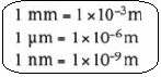
? Convert this measurement to µm by multiplying by 1 000.
50 x 1 000 = 50 000 µg
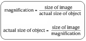
 ? Substitute into the equation:
- The magnification for this drawing:
- The length of the chloroplast:
? Measure the length of the image in mm (80 mm) and convert to µm ---> 80 000 µm.
? Calculate its real length:

5. Measuring cells using a graticule

Calibration: the conversion of graticule units into real units (mm, µm).
= 18 x 0.01 mm = 0.18 mm
= 180 µm
so 1 small eyepiece graticule marking = 180: 80 = 2.25 µm
- #75 Drawings
One of the questions in the exam is likely to involve drawing a specimen on a slide, using a microscope, or drawing from a photomicrograph (a photograph taking through a microscope). Making decisions about what to draw You might have to decide which...
- # 68 As Experimental Skills And Investigations
Almost one quarter of the total marks for your AS examination are for experimental skills and investigations. These are assessed on Paper 3, which is a practical examination. There is a total of 40 marks available on this Paper. Although the questions...
- #6 Summary Of Cell Structure
The basic unit of life, the cell, can be seen clearly only with the aid of microscopes.The light microscope uses light as a source of radiation, whereas the electron microscope uses electrons. The electron microscope has greater resolution (allows...
- #2.2. Cell Structure - Syllabus 2016
1.1 The microscope in cell studies1.2 Cells as the basic units of living organisms All organisms are composed of cells. Knowledge of their structure and function underpins much of biology. The fundamental differences between eukaryotic...
- Cell Organization, And Viewing Cells
Cells need mobility, sustainability, most cell sizes and shapes reflect their functions, and this is necessary for their homeostasis. Ways of seeing: Phase contrast takes advantage of variations in density within cells. the density variations alter certain...
Biology
#3. Microscopy
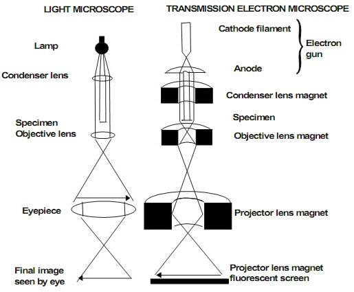
1. Light microscopes
- light rays pass through the specimen on a slide
- focused by an objective lens and an eyepiece lens.
- ---> magnified image of the specimen on the retina of your eye/screen/camera.
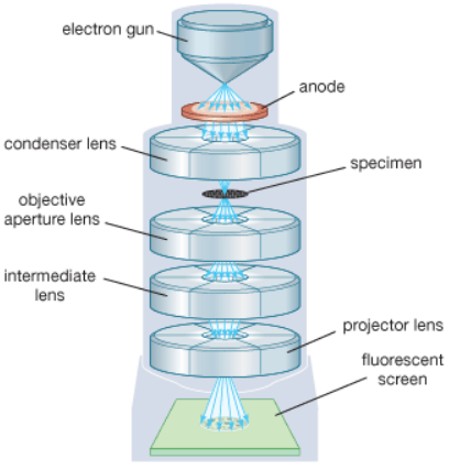
2. Electron microscopes
- uses beams of electrons
- specimen very thin, placed in a vacuum to allow electrons to pass through it.
- electrons are focused onto a screen/photographic film ---> magnified image of the specimen.
3. Magnification and Resolution
- Amount of magnification depends on the resolution of the microscope (ability to distinguish 2 objects as separate).
- The smaller the objects that can be distinguished --> the higher the resolution.
- wavelength: beam of electrons <<< light
- with electron microscope, we can see much more fine detail of a cell.
Units: millimetre, micrometre, nanometre
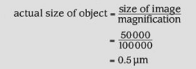
Work out the real size of an object knowing the magnification:
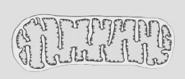
a. This drawing of a mitochondrion has been magnified 100 000 times.
? Use ruler to measure its length in mm (50 mm).
? Convert this measurement to µm by multiplying by 1 000.
50 x 1 000 = 50 000 µg
? Substitute into the equation:b. This is a the drawing of a chloroplast:
? Measure the length of the image in mm (80 mm) and convert to µm ---> 80 000 µm.
? Calculate its real length:
- Eyepiece graticule is a little scale bar placed in the eyepiece of light microscope.
- The graticule is marked off in 'graticule units'.
- Turn the eyepiece so that the graticule scale lies over the object: the width of one cell is 23 graticule units.
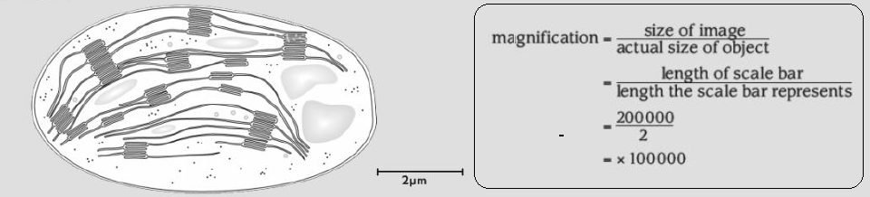
- use a special slide called a stage micrometer that is marked off in a tiny scale. The smallest markings are often 0.01 mm (10 µg) apart.
- Take the specimen off the stage or the microscope and replace it with the stage micrometer. Use the same objective lens.
- Line up the micrometer scale and the eyepiece graticule scale (by turning the eyepiece and moving the micrometer on the stage). Make sure that 2 large markings on each scale are lined up.
- The 50 mark (stage micrometer) is lined up with the 1.0 mark (eyepiece graticule).
- Work towards the right until you see another two lines lined up.
- The 68 mark (stage micrometer) is lined up with the 9.0 mark (eyepiece graticule). So you can say that:
= 18 x 0.01 mm = 0.18 mm
= 180 µm
so 1 small eyepiece graticule marking = 180: 80 = 2.25 µm
- The plant cell was 23 eyepiece graticule units long --> its real width is: 23 x 2.25 = 51.75 µm
Syllabus 2015: The microscope in cell studies Candidates should be able to: (a) [PA] use an eyepiece graticule and stage micrometer scale to measure cells and be familiar with units (millimetre, micrometre, nanometre) used in cell studies; (b) explain and distinguish between resolution and magnification, with reference to light microscopy and electron microscopy; (g) [PA] calculate linear magnification of drawings and photographs; (h) [PA] calculate actual sizes of specimens from drawings and photographs; |
Syllabus 2016: The microscope in cell studies An understanding of the principles of microscopy shows why light and electron microscopes have been essential in improving our knowledge of cells. a) compare the structure of typical animal and plant cells by making temporary preparations of live material and using photomicrographs b) calculate the linear magnifications of drawings, photomicrographs and electron micrographs c) use an eyepiece graticule and stage micrometer scale to measure cells and be familiar with units (millimetre, micrometre, nanometre) used in cell studies d) explain and distinguish between resolution and magnification, with reference to light microscopy and electron microscopy e) calculate actual sizes of specimens from drawings, photomicrographs and electron micrographs |
- #75 Drawings
One of the questions in the exam is likely to involve drawing a specimen on a slide, using a microscope, or drawing from a photomicrograph (a photograph taking through a microscope). Making decisions about what to draw You might have to decide which...
- # 68 As Experimental Skills And Investigations
Almost one quarter of the total marks for your AS examination are for experimental skills and investigations. These are assessed on Paper 3, which is a practical examination. There is a total of 40 marks available on this Paper. Although the questions...
- #6 Summary Of Cell Structure
The basic unit of life, the cell, can be seen clearly only with the aid of microscopes.The light microscope uses light as a source of radiation, whereas the electron microscope uses electrons. The electron microscope has greater resolution (allows...
- #2.2. Cell Structure - Syllabus 2016
1.1 The microscope in cell studies1.2 Cells as the basic units of living organisms All organisms are composed of cells. Knowledge of their structure and function underpins much of biology. The fundamental differences between eukaryotic...
- Cell Organization, And Viewing Cells
Cells need mobility, sustainability, most cell sizes and shapes reflect their functions, and this is necessary for their homeostasis. Ways of seeing: Phase contrast takes advantage of variations in density within cells. the density variations alter certain...